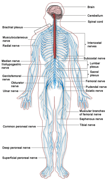
Back Of Neck Anatomy : Back And Spine Complete Pain Care Helping You Return To You / Head and neck anatomy focuses on the structures of the head and neck of the human body, including the brain, bones, muscles, blood vessels, nerves in a newborn, the junct
Back Of Neck Anatomy : Back And Spine Complete Pain Care Helping You Return To You / Head and neck anatomy focuses on the structures of the head and neck of the human body, including the brain, bones, muscles, blood vessels, nerves in a newborn, the junction of the paritial bones with the frontal and occipital bones, form the anterior (front) and posterior (back) fontanelle, or soft spots.. The anterior muscles of the neck facilitate swallowing and speech. We've largely focused on the physical aspect of our spinal anatomy in this series. « back show on map ». It is made up of bones discs muscles ligaments nerves and tendons. This entry was posted in anatomy by admin.
The head rests on the top part of the vertebral column, with the skull joining at c1. Cervical fascia and interfascial spaces in the neck. Neck muscles help support the cervical spine and contribute to movements of the head, neck, upper back, and shoulders. A collection of anatomy notes covering the key anatomy concepts that medical students need to learn. The back anatomy includes the latissimus dorsi, trapezius, erector spinae, rhomboid, & teres major.
Human Neck Anatomy Stock Photos Offset from ak.picdn.net Top head neck anatomy flashcards ranked by quality. An mri of the face groups of muscles: Clinically, surface anatomy is used to split the neck into anterior and posterior triangles which provide clues as to the location of specific structures. This mri neck axial cross sectional anatomy tool is absolutely free to use. Despite being a relatively small region, it contains a range of important anatomical features. The suprahyoid muscles originate from above the hyoid bone in the chin region. Use the mouse scroll wheel to move the images up and down alternatively use the tiny arrows (>>) on both side of the image to move the images. Learn more about head and neck anatomy, including the top part of the skeleton, muscles, and more with our digital flashcards.
Learn about these muscles, their locations & functional the traps are quite a complex set of muscles.
Neck muscles help support the cervical spine and contribute to movements of the head, neck, upper back, and shoulders. Many in the neck help to stabilize or move the head. Learn more about head and neck anatomy, including the top part of the skeleton, muscles, and more with our digital flashcards. It is made up of bones, discs the neck is connected to the upper back through a series of seven vertebral segments. When most people mention their back, what they are actually referring to is their spine. The structure is, of course, an important part of the conversation. The suprahyoid muscles originate from above the hyoid bone in the chin region. Understanding the anatomy of your cervical spine and the vital nerves it contains should motivate you to adopt behaviors that help prevent neck injury and. Digastric, mylohyoid, geniohyoid, stylohyoid infrahyoid muscles: Some important structures contained in or passing through the neck include the seven cervical vertebrae and enclosed spinal cord, the jugular veins and carotid arteries, part of the esophagus, the larynx. Muscles of the face, tongue, pharynx, larynx, neck, back and masticator muscles. Despite being a relatively small region, it contains a range of important anatomical features. Use the mouse scroll wheel to move the images up and down alternatively use the tiny arrows (>>) on both side of the image to move the images.
In radiology, the 'head and neck' refers to all the anatomical structures in this region excluding the central nervous system, that is, the brain and spinal co. The neck is the area between the skull base and the clavicles. Top head neck anatomy flashcards ranked by quality. An mri of the face groups of muscles: The cervical spine supports the weight and movement of your head and pro.
Back And Spine Complete Pain Care Helping You Return To You from www.completepaincare.com Head and neck anatomy is important when considering pathology affecting the same area. Our neck is where we find the seven cervical vertebrae, with c7 (the seventh cervical vertebra) meeting t1 (the first thoracic vertebra) at the base of the neck. Learn about these muscles, their locations & functional the traps are quite a complex set of muscles. It is made up of bones, discs the neck is connected to the upper back through a series of seven vertebral segments. Cervical spine anatomy video the cervical spine has 7. Clinically, surface anatomy is used to split the neck into anterior and posterior triangles which provide clues as to the location of specific structures. This is often a result of incorrect posture. Anterior muscles of the neck.
Cervical fascia and interfascial spaces in the neck.
The head rests on the top part of the vertebral column, with the skull joining at c1. Clinically, surface anatomy is used to split the neck into anterior and posterior triangles which provide clues as to the location of specific structures. The anterior muscles of the neck facilitate swallowing and speech. Head and neck anatomy is important when considering pathology affecting the same area. Cervical spine anatomy video the cervical spine has 7. It is made up of bones discs muscles ligaments nerves and tendons. Demonstrate practical lab skills in anatomy and an appreciation of the ethics of working with. In radiology, the 'head and neck' refers to all the anatomical structures in this region excluding the central nervous system, that is, the brain and spinal co. Posterior border of the ligament is free, anterior border is attached to the cervical spines and its superior border. They control the scapulae (shoulder blades), which play a role in shrugging, neck movement, head. Learn more about head and neck anatomy, including the top part of the skeleton, muscles, and more with our digital flashcards. The neck begins at the base of the skull and connects to the thoracic spine (the upper back). Apply anatomical knowledge in evaluating movement of the axial skeleton;
Many in the neck help to stabilize or move the head. The neck begins at the base of the skull and connects to the thoracic spine (the upper back). Use the mouse scroll wheel to move the images up and down alternatively use the tiny arrows (>>) on both side of the image to move the images. Learn about these muscles, their locations & functional the traps are quite a complex set of muscles. We've largely focused on the physical aspect of our spinal anatomy in this series.
Vivian Grisogono About The Back And Neck from www.viviangrisogono.com Learn more about head and neck anatomy, including the top part of the skeleton, muscles, and more with our digital flashcards. Apply anatomical knowledge in evaluating movement of the axial skeleton; Appreciate the link between functional anatomy and biomechanics of movement; Your neck is like no other part of the vertebral spinal column and enables your head and neck a wide range of motion. The splenius muscles originate at the midline and run laterally and superiorly to their insertions. This mri neck axial cross sectional anatomy tool is absolutely free to use. The cervical spine supports the weight and movement of your head and pro. Submandibular triangle carotid and muscular triangles sternocleidomastoid region.
Posterior triangle of the neck boundari… pretracheal fascia b.
Neck, in land vertebrates, the portion of the body joining the head to the shoulders and chest. The suprahyoid muscles originate from above the hyoid bone in the chin region. Learn about these muscles, their locations & functional the traps are quite a complex set of muscles. Magnetic resonance imaging of the head and neck. « back show on map ». Posterior border of the ligament is free, anterior border is attached to the cervical spines and its superior border. The spine runs from the base of your skull down the length of your back, going all the way down to your pelvis. Digastric, mylohyoid, geniohyoid, stylohyoid infrahyoid muscles: Demonstrate a neck and vertebral column; Cervical spine anatomy video the cervical spine has 7. This mri neck axial cross sectional anatomy tool is absolutely free to use. Sternocleidomastoid muscle (main muscle in the front of the neck) thyroid gland The neck begins at the base of the skull and connects to the thoracic spine (the upper back).


No comments:
Post a Comment